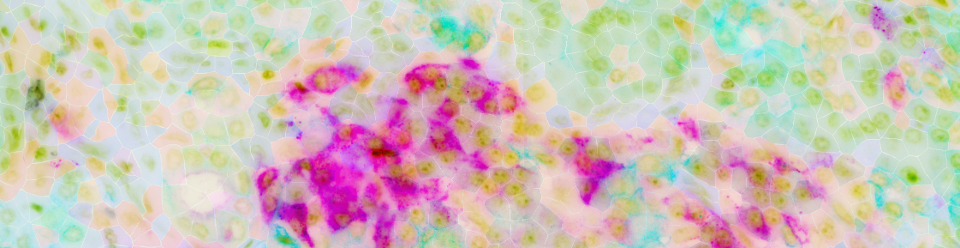
It is remarkable that evolution has produced organisms that behave as precisely balanced systems despite being built from an enormous number of distinct molecular components. A single mammalian cell assembles billions of protein molecules from tens of thousands of genes, diversified by splicing and other processing into a far larger set of distinct forms, alongside thousands of metabolites and a genome that is folded, marked and read differently in every cell type. Over the past two decades, genomic assays have gradually gained capacity to measure more and more of that complexity. Each generation of assays has widened the repertoire of observable molecular facets, from transcription to chromatin state, DNA methylation, or protein abundance. The assays have also increased the resolution, from bulk averages on tissue fragments to individual cells and now to individual molecules placed within the cell and within its tissue. Our group develops statistical and computational methods to interpret such genomic measurements, working in close collaboration with experimental groups to understand how normal tissues are built and how they break down in disease.
A key challenge is that living tissues are not merely collections of cells: a hepatocyte in isolation performs none of the functions we associate with a liver. Tissue functions, from immune defense to regeneration to the maintenance of an organ over a lifetime, are ensemble properties, arising from a sustained pattern of coordination among cells. Knowing the state of every cell in a tissue is necessary but not sufficient for understanding how tissues function, much as knowing the state of every component of an electronic circuit does not tell you what the circuit computes. Our group is interested in building predictive models of cell coordination: a quantitative description of the tissue as an integrated system rather than a catalogue of its parts. Such quantitative understanding requires answering several general questions:
What cells are influencing each other, and through what channels?
We currently lack robust methods to infer the communications taking place within a given tissue sample. What we need are models and inference methods with sufficient specificity to be validated experimentally, naming which cells act on which, through what channel, and to what effect. This is harder than it sounds, because signaling is not composed of independent interactions. Cells that receive signals also emit signals in return, so influences propagate through feedback loops, balancing many signals against one another. We would like to infer and model such feedback systems, ultimately to predict how they can be controlled. The immune compartment is the clearest example of why such models are needed: it integrates numerous signals, maintains balance around different functional set points, responds rapidly when needed, and plays a central role in a wide range of conditions, from the immunosuppressive tumor microenvironments to autoimmunity and aging.
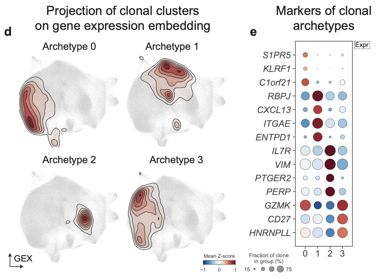
How does a cell process and integrate the external signals given its state and history?
Cellular influences unfold over time and different spatial scales. A cell integrates the impact of its immediate neighbors and extracellular matrix, diffusible signals from its local niche, as well as influences propagating across the whole organ. How these are balanced into a coherent response, and how that balance produces function at the level of the tissue, is largely unknown. The response also depends on the cell itself: earlier exposures can leave lasting traces, and cells from a common clonal origin often show shared predispositions. Modern lineage tracing methods can expose such behaviors. We are developing methods to follow clonal dynamics through development and adult tissue maintenance, and to characterize context- and history-dependent responses in hematopoiesis and in solid organs.
What regulates anatomical tissue structure at different scales?
Microanatomical structures combine to pattern tissue in a stereotypical manner across different scales. In the liver, hepatocytes are arranged into plates around sinusoids, sinusoids into lobules with a characteristic zonation, and lobules pattern the whole organ. At every scale such arrangements exhibit characteristic dimensions and composition, and no two instances are alike. These multi-scale patterns are progressively established during development, drift over time. It changes systematically with disease, for example under influence of chronic inflammation, fibrosis or invading tumors. We are developing approaches to quantify such multi-scale patterns, to study their variation across conditions, and to identify regulatory factors and mechanisms that underlie such patterning, ultimately aiming to arrive at dynamic models of tissue organization that are predictive rather than descriptive.
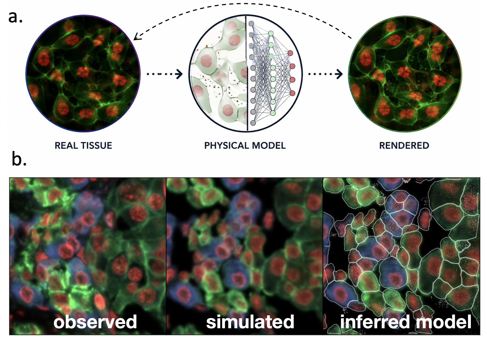
We envision that coordinated progress of experimental assays, statistical methods, AI models, and engineering of complex perturbations will allow to create models of cells and tissues that will become important instruments in their own right. Such models could then be used to answer questions about mechanisms, identify factors determining a given organ or disease phenotype, and to design more effective interventions.
We hope to advance quantitative understanding, and ultimately control, of how cells coordinate to establish and maintain functional tissues, and how these mechanisms fail in disease.
Our approach combines statistical modeling, machine learning and large-scale analysis of genomic, spatial and imaging data, alongside lineage tracing and perturbation experiments. All of this effort is performed in close collaboration with experimental groups, in the context of specific biological questions. The biology sets priorities for the methodological problems, and the experiments determine whether our solutions are effective.
Most of human diseases manifest on a tissue level. Auto-immune, inflammatory and fibrotic conditions, even tumor progression reflect failures of tissue homeostasis and protective mechanisms. Quantitative models of tissue function may allow to design more effective and targeted interventions at this level. A more immediate impact of our efforts is open-source analysis software that is used across academia and industry, enabling the studies of other groups.
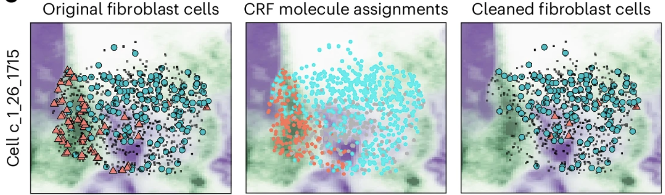
Mitchel, J., Gao, T., Petukhov, V., Cole, E., Kharchenko, PV. (2026). Impact and correction of segmentation errors in spatial transcriptomics. Nat Genet. 58(2):434-444
Mitchel, J., Gordon, MG., Perez, RK (...) Ye, CJ., Kharchenko, PV. (2025). Coordinated, multicellular patterns of transcriptional variation that stratify patient cohorts are revealed by tensor decomposition. Nat Biotechnol. 43(7):1192-1201
Gao, T., Soldatov, R., Sarkar, H (...) Loh, PR., Kharchenko, PV. (2023). Haplotype-aware analysis of somatic copy number variations from single-cell transcriptomes. Nat Biotechnol. 41(3):417-426
Petukhov, V., Xu, RJ., Soldatov, RA (...) Moffitt, JR., Kharchenko, PV. (2022). Cell segmentation in imaging-based spatial transcriptomics. Nat Biotechnol. 40(3):345-354
La Manno, G., Soldatov, R., Zeisel, A (...) Linnarsson, S., Kharchenko, PV. (2018). RNA velocity of single cells. Nature. 560(7719):494-498