Ion Imaging
The therapy of malignant cancer tumors using protons and carbon ions has become an established form of treatment in recent years due to its very precise controllability while sparing the surrounding tissue. With the commissioning of the MedAustron center, this therapy is now also available in Austria.
The exact location of the tumor must be known prior to treatment. This information is based on knowledge of the tissue and its composition, which the ions must pass through on their way to the tumor. Currently, this information is obtained from computed tomography using X-ray radiation. However, the ions used in particle therapy follow entirely different physical laws. They can be detected and their tracks can be recorded using techniques similar to those used for particles in high-energy physics. This informations allows the reconstruction of an image of the object passed by the ions.
To exploit this concept we are working on computed tomography with protons. We have built a prototype and are conducting tests with it within the framwork of non-clinical research at MedAustron. The following picture shows the concept of the system, which consists of spatially resolving tracking detectors. In between them, the object to be imaged is located. Behind this beam telescope, a calorimeter is placed to determine the residual particle energy after passing through the object.

The following image shows the actual measurement setup in the non-clinical irradiation room IR1 of MedAustron.

As a beam telescope, we use spatially resolving tracking detectors. Specifically, these are double-sided silicon strip sensors that were developed by us within the Belle-II SVD project. The front-end electronics is based on the APV25 readout chip, which in turn was designed for the CMS experiment and which we used extensively during our work for the CMS tracker. The picture on the right shows one of the six modules of this beam telescope.
The whole further readout chain, up to parts of the software, is again derived from the Belle-II experiment. This allows very efficient synergy, e.g. new components are tested in this setup first, before they are deployed in the Belle-II SVD. For example, a Gigabit Ethernet based readout was realized here for the first time.

To measure the energy of the ions after passing through the beam telescope and the object to be imaged, we use a sampling calorimeter. This uses 42 plastic scintillators, each with a SiPM (silicon photomultiplier) attached. This device, originally developed and called PRR30 for the Tera collaboration, had to be significantly refurbished by us. Thus, both the electronics (mainboard, FPGA firmware) and the readout software (from Labview to Python) were re-implemented.
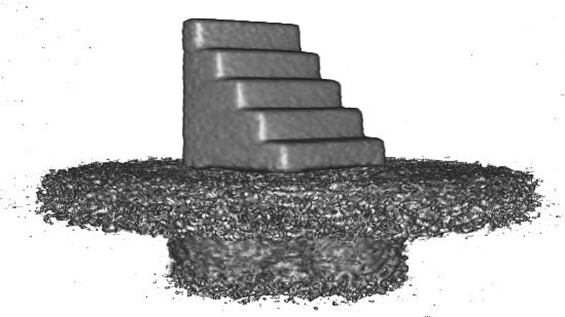
Since the project is pre-clinical, we are testing the concept by simulating and measuring phantoms. These are usually specially shaped bodies, like Catphan phantoms or simple cubes with a step profile. The picture on the left shows a photo of such a step cube next to a tracker module, while on the right is the reconstructed image of this cube.
Within the scope of this project, three PhD theses were written so far, covering the optimization of the tracking detectors, the energy measurement and image reconstruction. The last two ones were funded by the Austrian Research Promotion Agency FFG.