


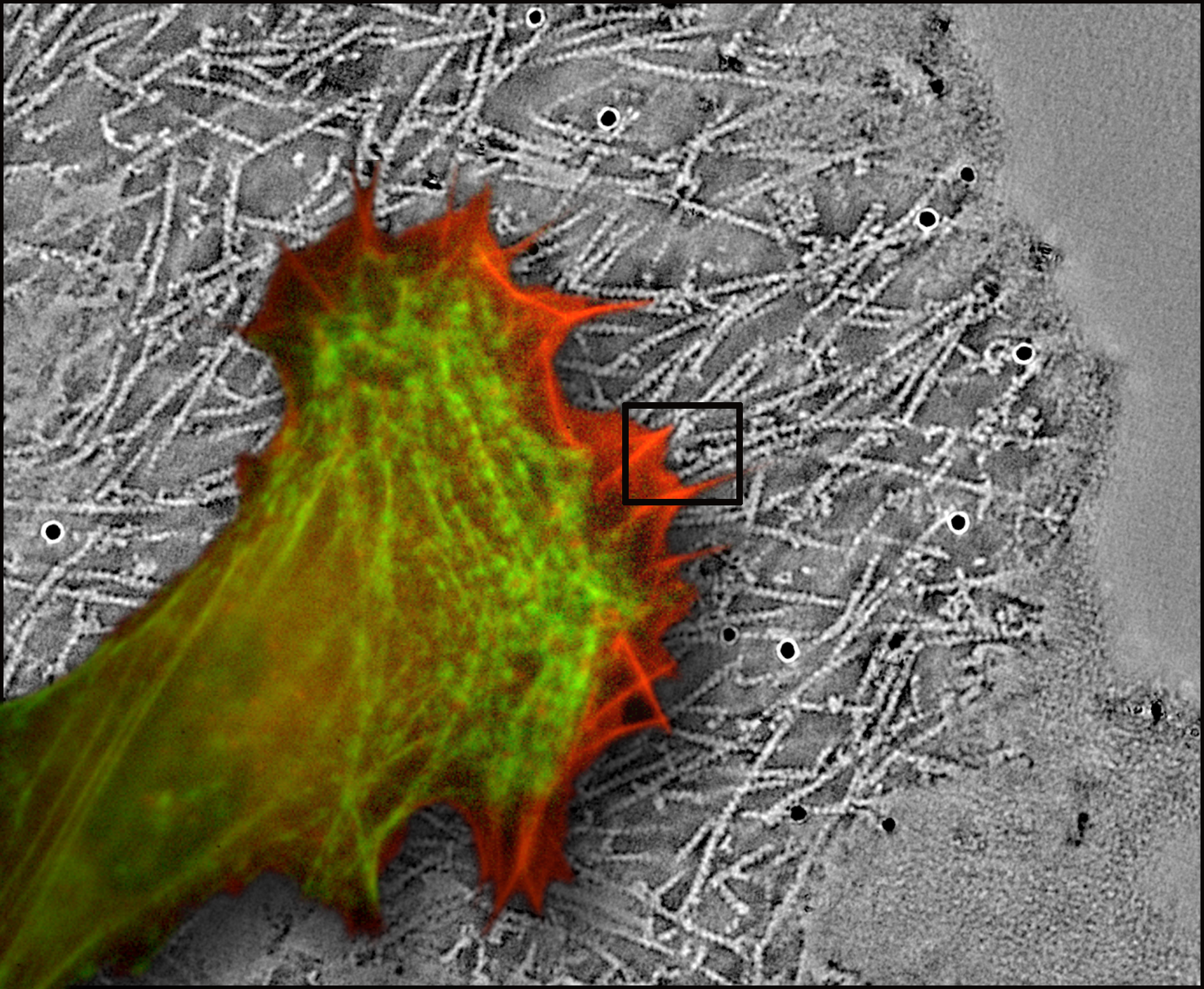
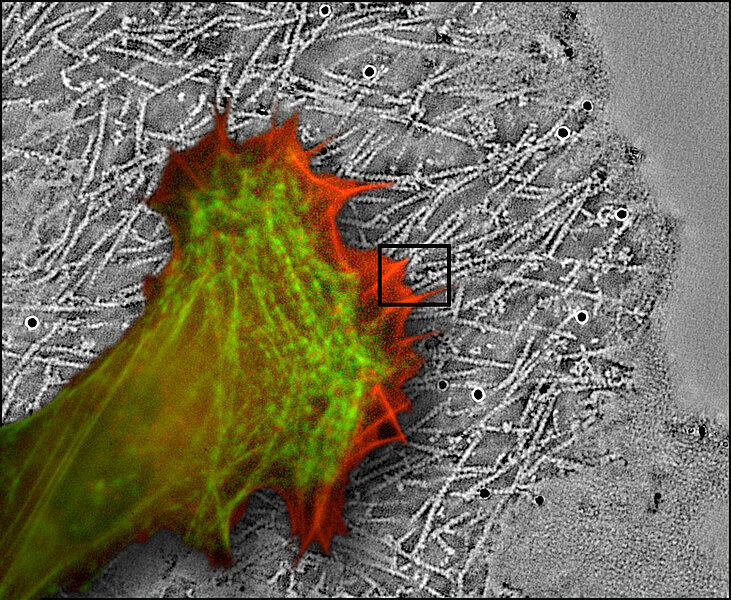
Numerous cells in our bodies are capable of active movement. They use an internal “motor” coupled with a cunning recycling system. Researchers from IMBA (Institute of Molecular Biotechnology) and IMP (Institute of Molecular Pathology) in Vienna have been able to literally freeze this process using cryo-electron tomography and to get a truthful picture of the cell’s internal propulsion system. Their findings, which challenge existing textbook models, make the cover-story of the current issue of the journal Nature Cell Biology.
Many different types of cells are able to migrate through surrounding tissue. Immune cells have to find and attack intruding pathogens, embryonic cells move towards their final destination during development, connective tissue cells close wounds. While movement of cells is essential for an organism to function properly, it can also be fatal, as in tumor cells disseminating from their origin and forming metastases in other parts of the body.
The cytoskeleton – a delicate structure within the cell
The cell’s internal “motor” is made up of numerous copies of the protein actin, lined up like beads on a string. The delicate actin-threads or “filaments” form an intricate network which acts as the cytoskeleton, a vitally important structure for the cell’s many tasks. Actin filaments have the ability to convey both pushing and pulling motions, which makes them extremely versatile. On the “front” end of the cell, actin molecules are clipped in one by one at the filament ends to push while at the rear end, they build filaments like in muscles to pull.
Unicellular organisms move rather clumsily by pushing their plasma forward like toothpaste being squeezed out of a tube. Migratory cells in higher organisms have to penetrate tissue and crawl between other cells that are tightly connected. They achieve this by forming finely shaped extensions (lamellipodia and filopodia) at the front end first and dragging the rest of the cell body behind.
British-Austrian cell biologist Vic Small, Senior Scientist at IMBA, has now been able to describe the components of the cell’s internal motor in their natural state. His data provide the structural basis for understanding actin-driven protrusion during cell migration. They also prove that the current textbook model is not correct and that some chapters will have to be rewritten.
Why actin filaments look like boiled spaghetti
The currently accepted model of how lamellipodia protrude includes actin filaments that branch like trees. The so-called dendritic nucleation model was supported by electron microscopic images that seemed to show quite clearly how the actin filaments branch. But cells subjected to electron microscopy had previously been treated rather harshly. The preparation of samples, which includes dehydration, fixation and coating with heavy metals, causes distortion and collapse of the delicate actin filaments in the cytoskeleton.
“What we have been looking at so far resembles a pile of soggy, boiled and squashed spaghetti”, says Vic Small. “We can now demonstrate that the dendritic appearance is a mere artifact and that apparent filament junctions are really just sites of filament overlap. Actin filaments are almost exclusively unbranched.”
Advanced technology unveils structures
To look at cells in their natural state, the scientists at IMBA used cryo-electron tomography, a combination of two methods which have merged into a powerful technology during recent years.
By using a cryo-electron microscope, the fixation process can be avoided. Instead, samples are deep-frozen and analyzed at minus 196 ̊C. Freezing the samples extremely rapidly, at a rate of -10 000 degrees per second, prevents the formation of ice crystals. Instead, the water vitrifies and thus conserves the internal structures in their natural state. Tomography makes it possible to generate three-dimensional images, allowing the unscrambling of densely packed structures in the cell.
The cryo-electron microscope is operated by Günter Resch, head of the service facility for electron microscopy at IMP-IMBA. His experience has been valuable in developing the technology further and has led to the construction of a device for rapidly freezing living cells, now commercially marketed by Leica Microsystems.
Understanding the motor that drives cell migration has implications beyond basic research. Vic Small: “We have to completely revise our opinion about cell movement. This will also help us understand how certain pathogens recruit the cytoskeleton to spread their infection.“ One of the follow-up projects will address the question how bacteria like Listeria penetrate cells and “hijack” the actin-motor for their own purposes.
****************
Original publication: “Electron tomography reveals unbranched networks of actin filaments in lamellipodia” by Edit Urban et al., Nature Cell Biology Vol 12, No 5, May 2010.
About Vic Small
John Victor Small was born in Orpington (UK) in 1944. He studied Physics at King’s College, London, where he obtained his doctorate in 1969. His research and teaching activities led him to the University of Aarhus (Denmark), the University of Melbourne (Australia) and Harvard University (USA). From 1977 to 2003, Vic Small was head of the Department of Cell Biology at the Institute of Molecular Biology of the Austrian Academy of Sciences, Salzburg. He was the institute’s director for a total of nine years. Since 2004, he is a Senior Scientist at the Institute of Molecular Biotechnology (IMBA) of the Austrian Academy of Sciences in Vienna.
About IMBA
The IMBA – Institute for Molecular Biotechnology of the Austrian Academy of Sciences – opened in 2003. It combines fundamental and applied research in the field of biomedicine. Interdisciplinary research groups address functional genetic questions, particularly those related to the origin of disease. The ultimate goal is to implement acquired knowledge into the development of innovative applications for prevention, diagnosis and treatment of disease.
About IMP - IMBA Research Center
A cooperation contract links the Institute of Molecular Biotechnology of the Austrian Academy of Sciences (IMBA) to the Research Institute of Molecular Pathology (IMP), which has operated since 1988 and is supported by Boehringer Ingelheim. Under the name of the “IMP – IMBA Research Center”, both institutes have access to a combined infrastructure in scientific and administrative areas. Together, the two institutes employ around 400 staff from 30 nations and are members of the Campus Vienna Biocenter.