


The field of structural biology deals with the molecular structure of molecules. The primary objective of this research area is to display the molecules as detailed as possible, in order to better understand how they work. "In structural biology, it is assumed that the shape of the molecules defines their function," says Thomas Marlovits *. Two very successful methods to determine structures, applied in structural biology so far, are the X-ray crystallography and NMR. For the X-ray crystallography sample preparation is needed, which means that crystals have to be grown out of a sample.
Over the last 30 years another method has evolved in parallel, the cryo-electron microscopy (cryo-EM). Here the molecules are put into a frozen state, pictures are taken and the three-dimensional atomic model is reconstructed. A big advantage of this method is that the sample does not require complex preparation beforehand. Recently, there were some exciting new developments, addressing the question how one can more efficiently extract signals from the often noisy images. On the one hand there were software innovations and, on the other hand, in the field of microscopy, new cameras were developed. "These new cameras are called direct electron detectors and allow the images to be sharper ", explains Thomas Marlovits.
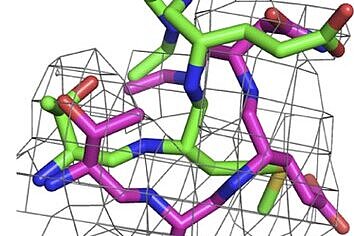
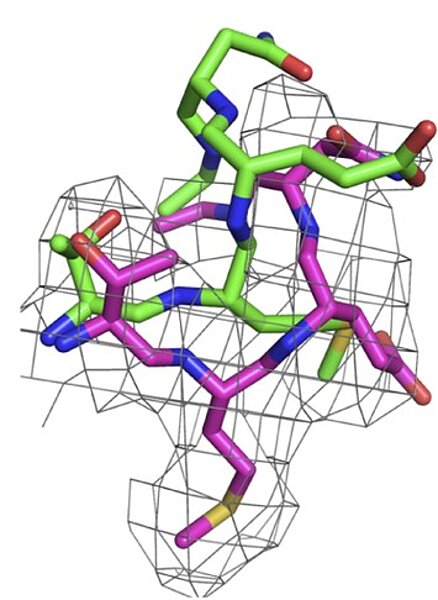
In their recent publication in Nature Methods a new approach has been presented. This procedure allows for even better and more accurate reconstructions of the atomic structures, using data from cryo-EM. A protocol was developed to reconstruct accurate atomic structures, even if the original data of cryo-EM is of lower resolution. It was found that the resolution, which is required to reconstruct an atomic structure can be at least 4.5 Å. To conduct this protocol a so-called electron density map is created, which is determined experimentally from electron microscopy images. Within the EM map, a X-ray crystallographic structure is placed and adjusted by the constraints provided by the EM map.
The presented method explains the mechanisms of molecules in atomic detail. For example, the best place for antibiotics to attach or why pathogenic bacteria often respond differently to antibiotics can be determined. "For questions like why some antibiotics work and others do not, the exact knowledge of the detailed structure is required. This refers not only to infectious diseases, but also to all other diseases where drugs are used. The better and in more detail we understand something, the better we can do something against it or for it," says Marlovits.
* Thomas Malovits is affiliated with the following organizations: Center for Structural Systems Biology (CSSB), University Medical Center Eppendorf-Hamburg (UKE), Hamburg, Germany, Deutsches Elektronen-Synchrotron (DESY), Hamburg, Germany, Institute of Molecular Biotechnology (IMBA), Austrian Academy of Sciences, Vienna, Austria, Research Institute of Molecular Pathology (IMP), Vienna, Austria.
Original publication
DiMaio, F., Song, Y., Li, X., Brunner, MJ., Xu, C., Conticello, V., Egelman, E., Marlovits, TC., Cheng, Y., Baker, D. (2015). Atomic-accuracy models from 4.5-Å cryo-electron microscopy data with density-guided iterative local refinement. Nat Methods. doi: 10.1038/nmeth.3286